


|

|
|
|

|
Search
05/25/2009
RESPONSES / COMMENTS (CLINICAL)
RE: Charcot Foot and Gout (Leonard Kuzmicki, DPM)
From: Multiple Respondents Ct scan and MRI are certainly helpful when diagnosing Charcot neuroarthropathy. This being said, a radiological report that suggests gout is just that, a suggestion. Degenerative changes seen in an MRI/CT scan are just one piece of the diagnostic puzzle. In order to accurately diagnose Charcot vs. an alternative degenerative process, a good history and physical exam is essential. Gerald Mauriello, Jr., DPM, MA, Philadelphia, PA, drmauriello@gmail.com I too have seen patients like this. However, the patients I had were being treated for hyperuricemia with uricosuric agents as well as Allopurinol for years. The patients also had extreme gouty attacks of which they were aware. It is possible that the patient has Charcot foot with underlying hyperuricemia. However, I am sure that they would have remembered extremely painful gout attacks, even with severe neuropathies. What is most likely is that this particular patient was not giving you a full medical history. Even with that being said, I would treat her as any other Charcot foot patient and consult a rheumatologist as these tophi can be reduced with low-dose Colchicine treatments. AS we know, the Charcot foot cannot. Judith Rubin, DPM, Houston, TX, jrubinfoot@aol.com It would be logical to expect that the same patient population which develops Charcot foot would be at higher than average risk for gout. Diabetes which has progressed to neuroarthropathy could be expected to take a significant toll on the kidneys as well. And these patients are also often on blood pressure meds which are known to increase risk of gout. But the sort of profound midfoot destruction that Dr. Kuzmicki describes is neuroarthropathy, not gout. Gout attacks are exquisitely painful. So, it is unlikely that a patient would walk on a foot during gout attacks enough to cause destruction of the navicular and cuneiforms with lateral dislocation of the metatarsals. Gout also destroys articular and peri-articular surfaces while Charcot neuroarthropathy is characterized by profound destruction which leads to foot collapse. If there is any question at all, a joint aspiration is indicated as that is necessary for a definitive diagnosis of gout. My rheumatologist wife says that it is rare, but gout can cause the extent of bone and joint destruction that Dr. Kuzmicki describes. This would most likely be accompanied by exquisite pain in the patient history. Richard Gosnay, DPM, Danbury, CT, glabroushead@gmail.com
Other messages in this thread:
09/07/2013
RESPONSES / COMMENTS (CLINICAL)
RE: Mueller-Weiss Syndrome
From: Joseph Borreggine, DPM
The standard of care for a failed conservative treatment plan for Mueller-Weiss syndrome is a solitary talonavicular joint fusion, or a full medial column fusion, or triple arthrodesis.
I have found that the medial column fusion seems to work best in these cases.
Joseph Borreggine, DPM, Charleston, IL, footfixr@consolidated.net
09/04/2013
RESPONSES / COMMENTS (CLINICAL) - PART 1A
RE: Bunion with Overlapping Toe
From: Michael B. DeBrule, DPM
Co, et al. looked at different interventions for 2nd toe transverse plane deformity in JFAS back in 2006. Syndacylization yielded the best alignment results compared to lateral anchor suture, flexor transfer, flexor plate repair etc. Therefore, I suggest: sew the 2nd and 3rd together, along with hammertoe corrections; 1st MTPJ athrodesis (more predictable toe alignment than with an Austin); and maybe a Weil 2nd osteotomy (depending on met length on x-rays).
On the other hand, the clinical picture seems more appropriate for an external metatarsal bar attached to an extra depth/custom shoe, custom foot orthotics, lift to correct limb length, periodic paring of the hyperkeratosis, or something conservative here. Consider discussing your proposed surgical intervention with the primary care doctor, physical therapist, and family. Would surgery truly improve her quality of life and help her walk better? Or would the patient be more likely to fall and sustain a hip fracture from wearing a CAM boot?
Michael B. DeBrule, DPM, Richfield, MN, innovativefootcare@gmail.com
09/04/2013
RESPONSES / COMMENTS (CLINICAL) - PART 1B
RE: Bunion With Overlapping 2nd Toe
From: Ed Cohen, DPM
A good point was raised about patient selection. A person with a drug or alcohol problem should be carefully screened and you might want to limit these type of patients to the more simple surgical procedures. It may be prudent not to take this case.
Amputation of the second toe should be avoided, as it will leave a space between the first and third toes, which will eventually cause the lateral deviation of the big toe and recurrence of the bunion. Also, an isolated second metatarsal head resection will ruin the...
Editor's note: Dr. Cohen's extended-length letter can be read here.
09/03/2013
RESPONSES / COMMENTS (CLINICAL)
RE: Bunion With Overlapping 2nd Toe
From: Hartley Miltchin, DPM, Philip J. Shapiro, DPM
This situation was similar to that of your patient.
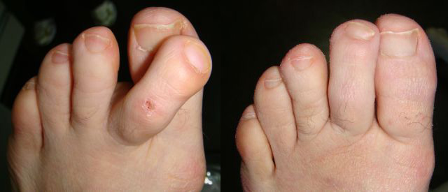 |
Pre- and post-op Photos |
I performed a modified Wilson/Silver/Akin with a diaphysectomy of the 2nd proximal phalanx. This procedure was accomplished with minimally invasive procedures. There were no sutures or internal fixation utilized. The foot was taped and a surgical shoe was used for six weeks, while the patient was fully ambulatory at all times. Extra strength Tylenol was used as needed without the need for narcotics.
Hartley Miltchin, DPM, Toronto, Canada, Doctortoe@aol.com
Why would anyone take this patient to surgery for an elective procedure if she cannot be trusted? As this woman’s ambulation is described as minimal in a nursing home environment, may I suggest a combination of periodic palliative care on the 2nd submetatarsal callus along with an accommodative soft shoe? If her lifestyle circumstances improve and she becomes trustworthy, then the surgical option can be revisited. Given her reported history, it would be a sound idea to have medical and/or psychiatric input prior to any elective invasive procedure.
Philip J. Shapiro, DPM, Ormond Beach, FL, pjsdpm@yahoo.com
09/02/2013
RESPONSES / COMMENTS (CLINICAL) - PART 1B
RE: Bunion With Overlapping 2nd Toe
From: Tip Sullivan, DPM, Michael J Marcus, DPM
Do you want to do foot surgery on a middle aged, non-compliant person who just happens to be an ex-addict? Is surgery the right thing to do? There is no doubt that surgical correction of this foot deformity is possible without the patient losing a toe. Whether you fuse the 1st MTPJ and syndactylize the second or do some other more proximal procedure, the question to ask here is one of patient selection for surgical intervention. If this unfortunate young gunshot victim is in a nursing home environment and is not walking much, why not get her some custom-molded shoes? If you do choose a surgical route for this patient, my advice would be “protect her from herself”— AK cast, knee flexed, toe pointed — and in a wheel chair until bone union.
Tip Sullivan, DPM, Jackson, MS, tsdefeet@msfootcenter.net
If symptomatic, I would perform an arthrodesis to the first MPJ together with a Weil osteotomy on the 2nd, arthrotomy/capsulotomy, or a plantar plate repair on the 2nd MPJ with a hammertoe fusion. These deformities are rigid secondary to neurologic etiology - gun shot/nerve injury. Nothing less than an arthrodesis would maintain the correction.
Michael J Marcus, DPM, Montebello/Irvine, CA, ftmed@aol.com
08/26/2013
RESPONSES / COMMENTS (CLINICAL)
RE: Traumatized Hallux Nail in 9 Year Old (David Gurvis, DPM)
From: Tip Sullivan, DPM
I guess that I am getting old or outdated or cheap, but let's simply look at the potential costs involved here for a known traumatic injury and the resultant digital enlargement and nail dystrophy.
X-ray - Let's just estimate $100
Rheumatology blood work—anywhere between $60 to $600 depending on tests ordered
MRI—national average -$1500
Office visit--???
So, let us say that potentially there could be a...
Editor's note: Dr. Sullivan's extended-length letter can be read here.
08/24/2013
RESPONSES / COMMENTS (CLINICAL) - PART 1
RE: Traumatized Hallux Nail in 9 Year Old (David E. Gurvis, DPM)
From: Dan Klein, DPM
Unfortunately, when a heavy object drops on a toe involving the toenail, permanent soft tissue changes will occur. In this case, a young person's toe was involved with apparent soft tissue changes. I believe these changes are permanent, but over time as this person grows, the changes will become less obvious. With regard to the toenail, the growth plate may also have been permanently damaged, producing a permanent change. The analogy I have used in practice is to compare the trauma with a bowling ball dropping on a tomato. The tomato will never look the same.
Dan Klein, DPM, Fort Smith, AR, toefixer@aol.com
08/24/2013
RESPONSES / COMMENTS (CLINICAL) - PART 2
RE: Wart Treatments (Dusty McCourt, DPM)
From: Paul Kesselman, DPM
Over the last few years, I have stopped almost all surgical and office-based chemical care for verrucae (even mosaic). If a few treatments of OTC chemcals fail to make any headway, I send the patient to the pharmacy with an Rx for 1 ml of Candida antigen. The cost is '$125 and will last approximately ten treatments. The candida is mixed 0.1 cc, with an equal amount of lidocaine (with or without epi) and I inject this directly into the verrucae. As this may be painful, the area must first be anesthetized with lidocaine/epinephrine.
With mosaic verrucae, I choose the largest "mother" wart. The treated area is re-evaluated every two to three weeks. I have rarely had to inject a verruca more than three times. Your patients (and you) will be amazed at the results. Two little known articles can be read on this subject and may be found online at: altoonafp.org/full.htm and podiatryinstitute.com/pdfs/Update_2012/2012_05.pdf
Prior to embarking on this regimen, one should obtain a good medical history and be sure your patient has no history of yeast sensitivities or previous fungal infections.
Paul Kesselman, DPM, Woodside NY, drkesselmandpm1@hotmail.com
08/05/2013
RESPONSES / COMMENTS (CLINICAL)
RE: Cyclist with Bilateral Forefoot Pain (Mark Aldrich, DPM)
From: Steven E. Tager, DPM
I've seen this quite a few times in cyclists. It's likely a compression neuropathy secondary to excessive rearfoot pronation. As the rearfoot pronates, the forefoot abducts into the lateral wall of the shoe which is locked in place by the clip. The foot swells as its irritated and the compression becomes more significant. Wider shoes are of some value, but in my experience, addressing the excessive pronation with a quality rearfoot posted orthotic eliminates the etiology, and the compression neuropathy subsides.
Steven E. Tager, DPM, Cupertino, CA, setdpm44@yahoo.com
07/29/2013
RESPONSES / COMMENTS (CLINICAL) - PART 1A (CLOSED)
RE: Chronic Pruritic Lesion (Arthur Gudeon, DPM)
From: Don Steinfeld, DPM
That looks like lichen simplex chronicus to me. Originally a pruritic lesion, the skin becomes indurated with exaggerated skin lines due to repetitive excoriation. This is an example of neuro-dermatitis. Try an Unna boot to physically prevent access to the site.
Don Steinfeld, DPM, Farmingdale, NJ, footdrdon@aol.com
07/29/2013
RESPONSES / COMMENTS (CLINICAL) - PART 1B (CLOSED)
RE: Chronic Pruritic Lesion (Arthur Gudeon, DPM)
From: Art Gudeon, DPM
Thanks for your responses; they’re much appreciated. As it turns out, Bryan Markinson, DPM, my “go-to podiatric dermatologist,” agreed with the consensus that the condition was NOT psoriasis, but lichen simplex chronicus. He advised treating this condition with clobetasol 0.05% bid (basically the same potency as the betamethasone dipropionate 0.05% ung., bid, that I’d already prescribed). I can alter the Rx later if necessary, and also add Benadryl prn (although she says the pruritis is minimal right now).
Just to clarify the biopsy situation - the reason she preferred not to have it on her first visit was due to a swimming competition “final” later that day, but said she’d want it if the response wasn’t proving satisfactory over a “reasonable” period of time. She was aware she’d have to D/C treatment 48-72 hours prior to the biopsy if so.
Art Gudeon, DPM, Rego Park, NY, afootdoc@hotmail.com
07/08/2013
RESPONSES / COMMENTS (CLINICAL) - PART 1a
RE: Surgical Intervention in a 12 Year Old with Flat Foot
From: Peter Bregman, DPM
The only thing I would do before making a surgical decision is do a neutral position x-ray and determine if a Cotton is really needed. Having said that, I recommend a Gastroc recession and a Hyprocure subtalar stent. Then add a Cotton, if needed, and then an Evans, if needed. In most cases, the Evans is not needed, but you can always tell the patient that they may need another procedure down the road.
If the clinical picture shows significant calcaneal valgus, then consider a calcaneal slide osteotomy instead of the Evans. I think Calc slide osteotomies are over-utilized because surgeons are not using enough arthroereisis implants which often obviate the need for these osteotomies.
Peter Bregman, DPM, Las Vegas, NV, drbregman@gmail.com
07/08/2013
RESPONSES / COMMENTS (CLINICAL) - PART 1B
RE: Surgical Intervention in a 12 Year Old with Flat Foot
From: Vincent Gramuglia, DPM, Mort Wittenberg, DPM
If there is no associated peroneal spasm, I would consider the TAL along with calcaneal osteotomy and plantarflexory medial column procedure like a Cotton or a Lapidus. Your philosophy should be to increase varus of rear foot and increase valgus of forefoot as taught by Dr. Dennis Shavelson.
Vincent Gramuglia, DPM, Bronx, NY, a2onpar3@optonline.net
In the “old days”, a TAL, followed with well-made custom orthoses, and at least a year of follow-up, would be done before all those extra surgeries would be considered.
Mort Wittenberg, DPM, Evans, GA, mwittenberg3@comcast.net
04/30/2013
RESPONSES / COMMENTS (CLINICAL) - PART 1
RE: Wartabater (Greg Caringi, DPM)
From: Art Hatfield, DPM
Anyone who has been in practice for more than a few months knows that NOTHING is "virtually always 100% successful." Is the word "virtually" a subtle disclaimer? If the manufacturer is that confident, ask for 10 free treatments to evaluate how it does in your practice. If it is 100% successful as they claim, they should have no problem with your request. We all know that the "wart" is a dermal manifestation of a viral infection. Until a vaccine against the human papilloma virus is available, we as a human race, will be cursed with warts.
Art Hatfield, DPM, Long Beach, CA, afootjob@juno.com
04/30/2013
RESPONSES / COMMENTS (CLINICAL) - PART 2
RE: Non-Specific Bone Marrow Edema Syndrome
From: Erik Kenyon, DPM, Eric J. Roberts, DPM
Sometimes, the non-specific marrow edema in pediatric patients is normal. It is believed to be as a result of transition of the bone marrow from infancy to adulthood.
Here is a reference: Shabshin N, et. al. High-signal T2 changes in the bone marrow of the foot and ankle in children: red marrow or traumatic changes? Pediatr Radiol. 2006, 36: 670-676.
Erik Kenyon, DPM, Modesto, CA, Erik.Kenyon@kp.org
Note that the non-specific bone marrow edema seen on an MRI in children that are the age of your patient is a normal finding due to the hematopoetic cells. The pathology you are looking for is unlikely this non-specific finding. Over 50% of children have this finding bilaterally and is normal in growing bones. Focus on the ankle instability and continue your treatment.
Pal CR, Tasker AD, Ostlere SJ, et al. Heterogeneous signal in bone marrow on MRI of children's feet: a normal finding? Skeletal Radiol. 1999;28 (5): 274-8.
Eric J. Roberts, DPM, Tampa, FL, lordertz@gmail.com
04/06/2013
RESPONSES / COMMENTS (CLINICAL) - PART 2A
RE: Recalcitrant Heel Pain (Loren J Miller, DPM)
From: Paul R. Scherer, DPM, Mark Hinkes, DPM
I suggest that you work the patient up for sero-positive or sero-negative arthritis. The following diseases have been well documented as having unilateral persistent heel pain as the presenting symptom: sero-negative (HLAB27 positive) include ankylosing spondylitis, reactive arthritis (formerly Reiters), enteropathic spondylitis, psoriatic arthritis, anterior uveitis. Seropositive include rheumatoid arthritis, systemic lupus, and Sjogren’s syndrome. Custom orthotics sometimes don’t work. Who would have thought!
Paul R. Scherer, DPM, San Francisco, CA, pscherer@prolab-usa.com
When does the pain occur? Does the patient have AM pain upon arising or neuritic type pain in the afternoon? Does the patient have an unequal limb length? Is there pain to the calcaneus with medial/lateral compression? Is there a positive lamp cord sign to the medial calcaneal nerve? How about a bone scan of the calcaneus to R/O sub-clinical stress fracture? Does that patient have any history of lumbar spine trauma/surgery? Does the patient have any history of DVT? Have you tried a bone stimulator or casting the patient to be NWB for 6 weeks? Have you run any labs? ANA sed rate, crp, uric acid, bpm, rheumatoid factor?
Mark Hinkes, DPM, Murfreesboro, TN, drhinkes@gmail.com
01/16/2013
RESPONSES / COMMENTS (CLINICAL) - PART 2
RE: The Self-Esteem of Podiatry 2013 (Gino Scartozzi, DPM)
From: Jack Sasiene, DPM
Dr. Scartozzi states: "The solution is quite simple, the decision to provide treatment procedures for a patient should not be contingent on where the source of payment will be coming from. It is not the practitioner who is responsible to routinely write-off services that are not covered by insurance carriers." This is my point exactly. Medical care in this country has been dictated by the insurance carriers. Medicare sets our fee payments, private insurance uses it, and have brainwashed patients into the concept that everything is covered and we are the guilty parties when we aren't. Why be put in the middle of that?
I wonder what "smart business doctors" like Jon Hultman, David Helfman, and Hal Ornstein would say about...
Editor's note: Dr. Sasiene's extended-length letter can be read here.
12/24/2012
RESPONSES / COMMENTS (CLINICAL)
RE: Displaced MBA Implant (Neil Levin, DPM)
From: Darryl Burns, DPM
I had a similar patient, a 22 year old male who sprained his ankle, and the implant rotated 90 degrees. It blocked the motion of the STJ in a fixed varus position. He had an asymptomatic MBA implant in the other foot, both of approximtaely 15 years. Due to the pain, he elected to have the implant removed. In addition, he wanted the other implant removed at the same time.
The removal of the implant simply requires an incision over the sinus tarsi and with some blunt dissection, the implant will be easily identified. Using a 3.5 hex head screwdriver, it can be inserted into the implant and unscrewed. I followed up with orthotics that I use for PT tendon dysfunction. I have a satisfied patient. There are some arthritic changes on the displaced implant side (noted on x-ray), but no clinical symptoms.
Darryl Burns, DPM, Monterey, CA, darrylburns@msn.com
12/17/2012
RESPONSES / COMMENTS (CLINICAL)
RE: Tibial Sesamoid Fracture (Jeffrey Kass, DPM)
From: Barry Mullen, DPM, Bhavin Mehta, DPM
In regard to Dr. Kass' comment on injecting a "liquid bone" under ultrasound guidance and then appling a bone stimulator for a sesamoid fracture, the question that arises is coverage for these treatments by insurance. I think the proposed idea is a good conservative approach to try to heal the fracture, but I'm not sure about whether or not this would be covered.
Bhavin Mehta, DPM, Newnan, GA, tarheel72@hotmail.com
I admit treatment outcomes for sesamoid fractures can be perplexing, and in some cases, less than ideal. Physiologic reasons exist. Previous literature discusses off-loading, PT, injections, and removal for symptomatic non-unions. I once read about an ORIF with small cortical screw placement across a sesamoid fracture that was tried, but never read any long-term follow up on its success, and whether any other similar attempts were tried; so, I naturally assumed the outcome was not very good, and the surgeon abandoned that particular approach.
From personal anecdotal experience, the overwhelming majority of sesamoid fractures do not heal, yet their non-unions tend to...
Editor's note: Dr. Mullen's extended-length letter can be read here.
12/15/2012
RESPONSES / COMMENTS (CLINICAL)
RE: Tibial Sesamoid Fracture (Terry Nayfa, DPM)
From: Jeffrey Kass, DPM
I would like to throw out to the readers a possible treatment plan for the tibial sesamoid fracture. This is not something that I have tried, but it is something I considered a few months back on a sesamoid fracture that presented in my office. I am presenting it with the hopes of constructive critique in both the positive or negative.
What if one injected one of the "liquid bone" products on the market under ultrasound guidance after needling the region between the bony fragments. The intent would be to bridge the fragmented parts together. This would be followed by bone stimulation and off-weight-bearing. If it didn't work - at least you tried. If it did work, well you saved the patient from being cut open, which in an athlete could have a deleterious effect.
Jeffrey Kass, DPM, Forest Hills, NY, jeffckass@aol.com
12/12/2012
RESPONSES / COMMENTS (CLINICAL) - PART 1B
RE: Tibial Sesamoid Fracture (Terry Nayfa, DPM)
From: Elliot Udell, DPM
We have had great success in treating fractures of the sesamoids using the Exogen bone stimulator. The trick, however, is to maximally off-load the area in order to allow for healing. To this end, you need to have a good working relationship with the company that makes your custom orthotics and have them design a device that will take weight off of the first met head.
After the orthotic arrives, mark the area of the first met head and have the patient walk on the orthotic to make sure the correction is in the correct location. You may have to supplement even the best-made orthotic with layers of felt. Let the patient know that it may not be the most comfortable device, but unless the weight is off-loaded from the area, healing of the fracture will be impaired.
Elliot Udell, DPM, Hicksville, NY, Elliotu@aol.com
12/12/2012
RESPONSES / COMMENTS (CLINICAL) - PART 1A
RE: Tibial Sesamoid Fracture (Terry Nayfa, DPM)
From: Keith L. Gurnick, DPM
1) For now, forget the orthotics. Try taping the foot/toe with a dancer's type pad and a turf toe type taping to off-load the sesamoid and limit 1st MPJ motion (daily and in sports).
2) Order an MRI to make sure the involvement is at the proximal sesamoid bone only.
3) Be certain you do not have any involvement of the flexor hallucis longus (tendinitis)
4) If you perform surgery and remove only the proximal sesamoid, make certain to place one or two "stay" sutures in the plantar medial ligament to tighten up the void.
5) After the patient heals from the surgery and returns to activity, continue taping for 6-8 weeks.
Note: From the limited information and zoomed in x-ray, it appears he has a rounded shape to the first metatarsal head and a slight hallux adductus. Removing the entire sesamoid (both pieces) in such a young active athlete could hasten the development of a hallux valgus as he ages. He should be informed of this before surgery.
Keith L. Gurnick, DPM, Los Angeles, CA, keithgrnk@aol.com
12/11/2012
RESPONSES / COMMENTS (CLINICAL) - PART !B
RE: A1c and Elective Foot Surgery (Allen Jacobs, DPM)
From: Elliot Udell, DPM
Our primary concern in performing surgery on patients with diabetes is whether they will uneventfully heal after the surgery. Abnormally elevated blood glucose levels over a long period of time, which can be measured by testing the hemaglobin A1c, may be one way of deciding whether to exclude elective surgery on a patient with diabetes. Two other causes of potential post-operative problems in these patients are poor pedal vascularity and severe peripheral neuropathy. Hence, prior to considering doing any elective procedures on these patients, a good arterial vascular exam including digital studies should be performed.
In addition, neurological testing, which would include rudimentary tests such as vibratory sensation and light touch testing, can be in done. In addition, good nerve conduction studies should be considered. Many podiatric physicians own the proper equipment and do these studies regularly. For those of us who do not, there are mobile vascular and nerve conduction testing companies that will come to our offices and perform these tests and provide interpretations of the tests by appropriate specialists.
Elliot Udell, DPM, Hicksville, NY, Elliotu@aol.com
12/10/2012
RESPONSES / COMMENTS (CLINICAL) - PART 1B
RE: A1c and Elective Foot Surgery (Allen Jacobs, DPM)
From: Tip Sullivan, DPM
Dr. Jacobs has opened a very appropriate topic for discussion. I am not aware of any papers that specifically relate A1c to foot surgery success, elective or otherwise. The first thing that I asked myself after considering his question was: What is “elective” foot surgery? Certainly, we all agree that the current trend in “cosmetic” foot surgery is “elective.” I think the line one draws between elective surgery and necessary surgery can get grey.
Perhaps the terms emergent and non-emergent surgery would be...
Editor's note: Dr. Sullivan's extended-length letter can be read here.
12/08/2012
RESPONSES / COMMENTS (CLINICAL) - PART 1 A
RE: A1c and Elective Foot Surgery (Allen Jacobs, DPM)
From: Pat Caputo, DPM
We all know that the hemoglobin A1C test measures the average blood glucose control for the past 2 to 3 months and is a good general measure of glycemic control during that period. Due to the nature and definition of glycosylated hemoglobin, it only needs to be performed 3 times per year. I am not so sure it should be considered or classified as standard of care to order a hemoglobin A1c prior to all elective foot surgery, unless it hasn’t been checked in over 3 months. It is, however, a very useful tool in predicting increased risk. Studies have shown that “Elevated pre-operative hemoglobin A1c level is predictive of adverse events after coronary artery bypass surgery” (Halkos, M et al., Journal of Thoracic and Cardiovascular Surgery. 2008. 136(3) 631-640.)
The surgeon has to determine how extensive the planned surgery is and measure all other co-morbidities and factors (obesity, smoking, patient compliance, etc.). A HgA1c level of >9% represents an obvious high level of risk that I wouldn’t want to have my diabetic patient exposed to. A target A1c of under 7% is the most appropriate. As the A1c increases, so does the risk of complications. Like most everything we do, your medical decision is how much risk is worth the benefit.
Pat Caputo, DPM, Holmdel, NJ, capstops@aol.com
|
| |

|
|
|